 |
| Dark purple form of P. forskalii. Photo credit: Ria Tan. |
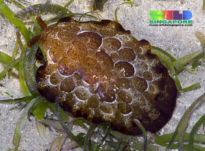 |
| Intermediate brown form of P. forskalii. Photo credit: Ria Tan. |
Table of Contents
Pleurobranchus forskalii (Rüppell & Leuckart, 1828) or Forskal's side-gilled slug (from Greek: pleurón = side or rib; branchion = a gill) belongs to a group of marine gastropods which possess a single gill on the right side of their body. Members of this group are capable of attaining large sizes (≥ 30 cm in some species), and are also often brightly coloured with striking patterns, making them easily one of the more charismatic intertidal organisms. P. forskalii is very variable in colour, with individuals ranging from dark purple (pictured above; left) to light brown. An intermediate brown form is pictured on the right. P. forskalli possesses important biologically active compounds which may be effective in the treatment of some cancers.
Inspiration for This Page
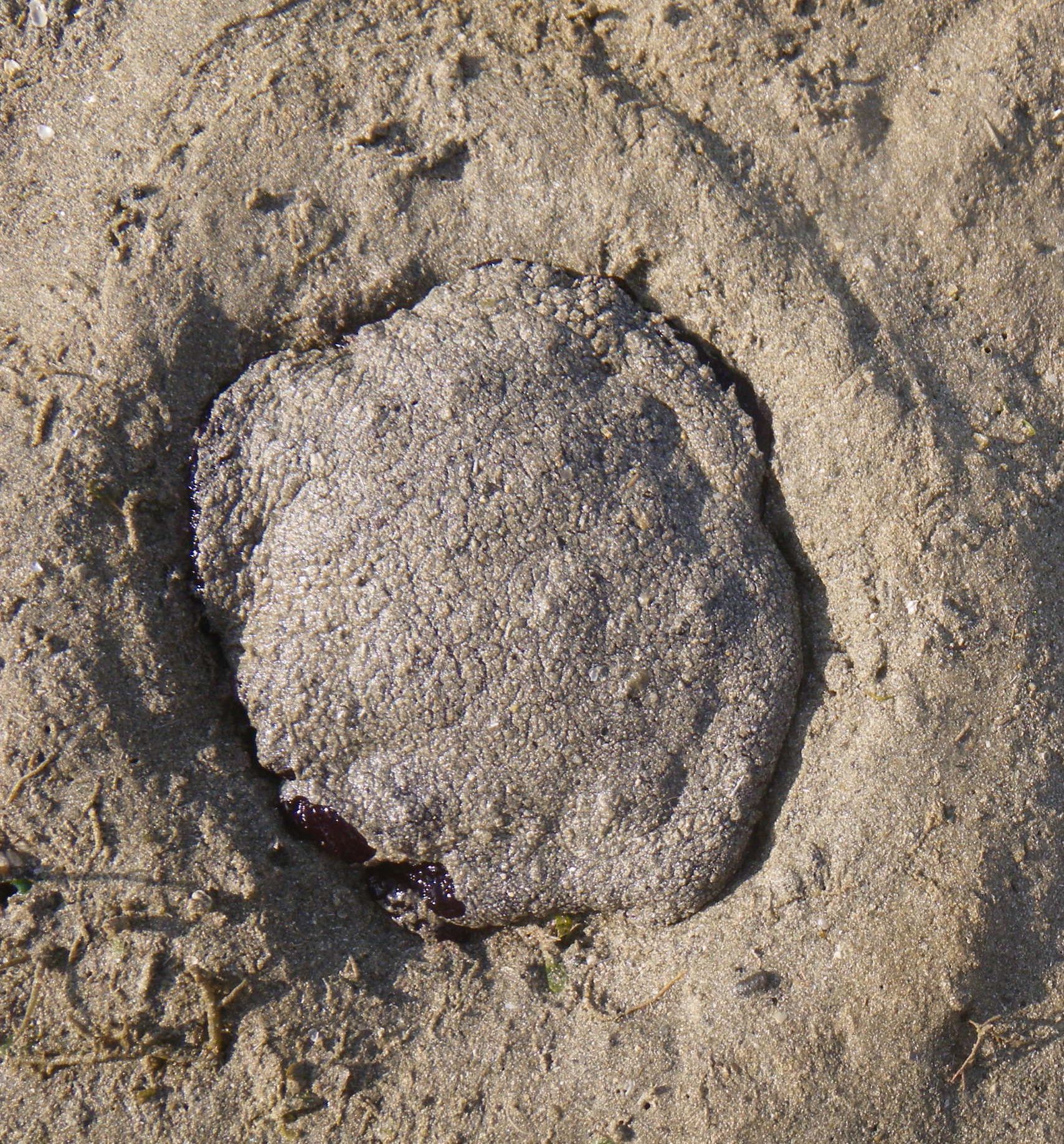 |
| Stranded P. forskalii during low tide. Own photo (Henrietta Woo). |
General layout
The page opens with pictures of P. forskalii illustrating its variable colour forms and then delves into the biology as well as other aspects of the animal such as its behaviour and uses. More technical information such as its phylogeny can be found towards the end, which may appeal to those interested in taxonomy. Technical jargon has been kept to a minimum, and where used, links to external websites which explain the terms have been included to aid the understanding of the reader. In addition, most photos are linked to the original should readers wish to view the original.The Many Colours of Forskal's Side-gilled Slug

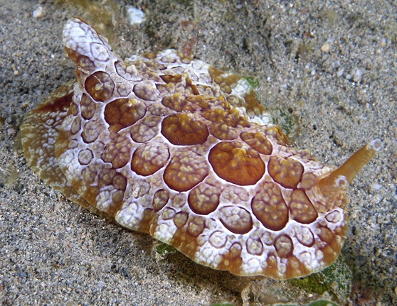 |
| Photo credit: Nick Hobgood. |
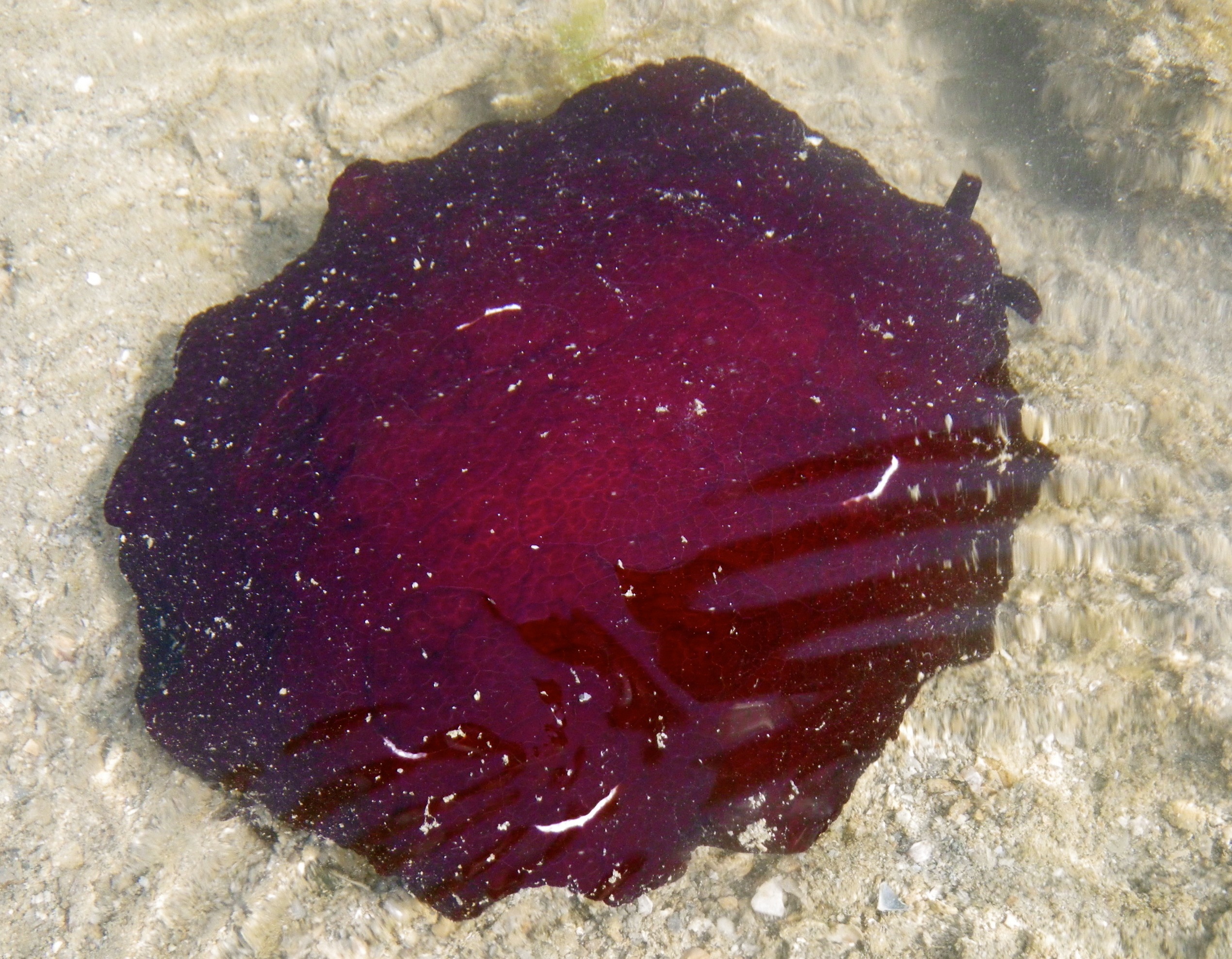 |
| Own photo (Henrietta Woo). |
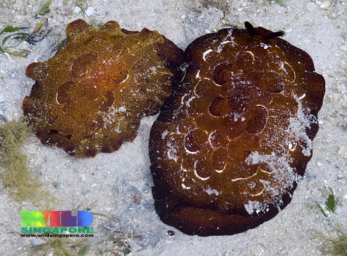 |
| Photo credit: Ria Tan. |
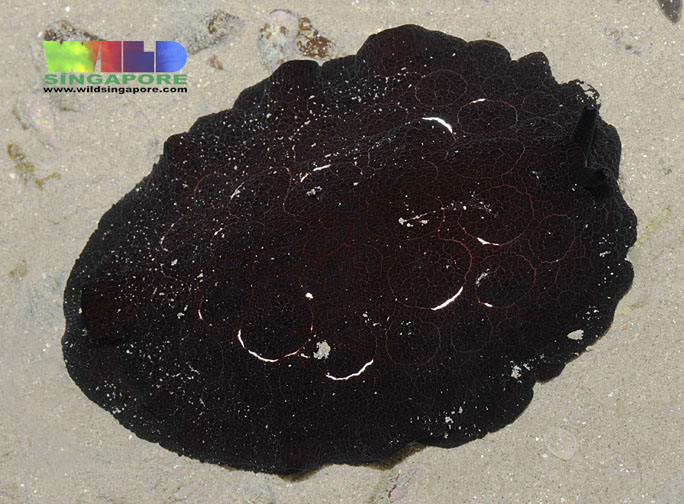 |
| Photo credit: Ria Tan. |

Hey, I thought I saw a...
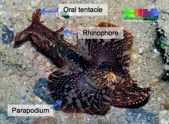
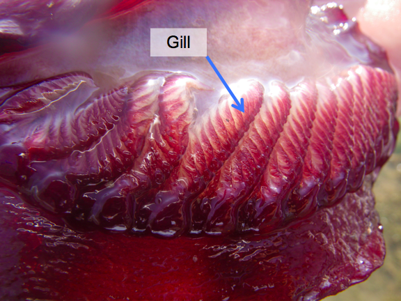
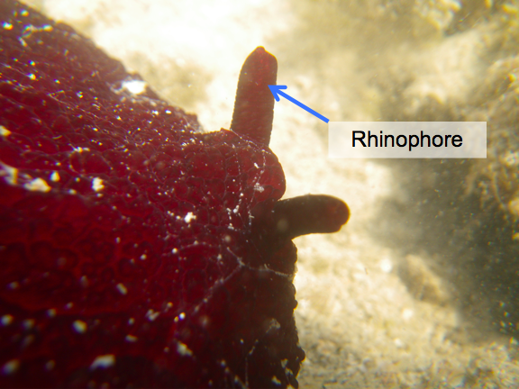
To the casual observer, side-gilled slugs may sometimes be mistaken for other sea slugs including nudibranchs and sea hares. The surest way to tell them apart is to look for the following distinguishing characteristics (homologous structures have also been marked out):
..Side-gilled Slug
 |
 |
| Pleurobranchus forskalii. Own photo (Henrietta Woo) with modifications. |
P. forskalii. Own photo (Henrietta Woo) with modifications. |
- As their name implies, side-gilled slugs possess a gill only on one side of their body (i.e. the right side), between the mantle and foot (see 'Morphology' below).
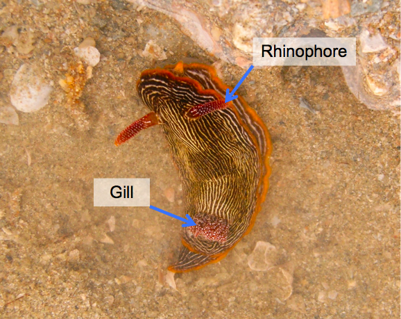
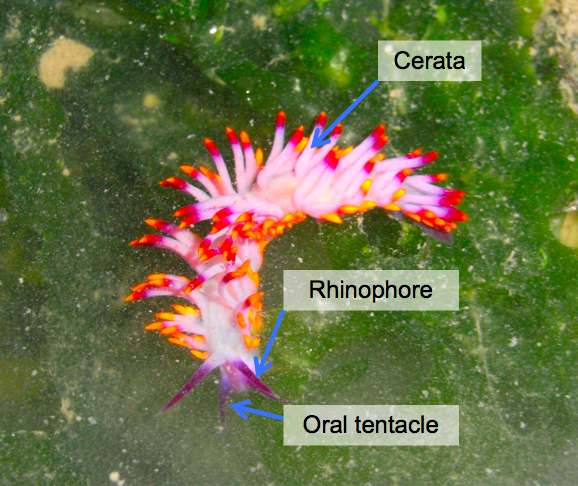
..Nudibranch
 |
 |
| Chromodoris lineolata. Own photo (Henrietta Woo) with modifications. More information at http://www.wildsingapore.com/wildfacts/mollusca/slugs/nudibranchia/lineolata.htm. |
Cuthona sibogae. Own photo (Henrietta Woo) with modifications. More information at http://www.wildsingapore.com/wildfacts/mollusca/slugs/nudibranchia/sibogae.htm. |
- Many nudibranchs have an exposed gill on the dorsal surface of their body, located posteriorly.
- Others possess outgrowths on their body known as cerata (singular: ceras).
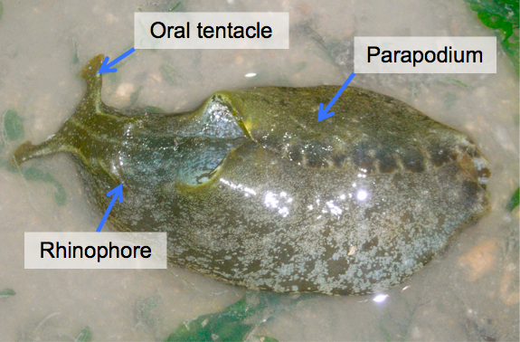
..Sea Hare
 |
|
| Aplysia extraordinaria. Image courtesy of Ria Tan (with modifications). More information at http://www.wildsingapore.com/wildfacts/mollusca/slugs/anaspidae/extraordinaria.htm. |
Aplysia species. Own photo (Henrietta Woo) with modifications. More information at http://www.wildsingapore.com/wildfacts/mollusca/slugs/anaspidae/spotted.htm. |
- Sea hares have prominent oral tentacles as well as rhinophores. When erect, the latter supposedly resemble ears of a hare/rabbit, giving them their name.
- They possess large parapodia (singular: parapodium) that may be used for swimming.
Two's Company, Three's a Crowd
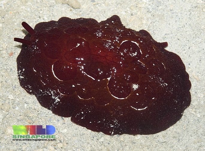
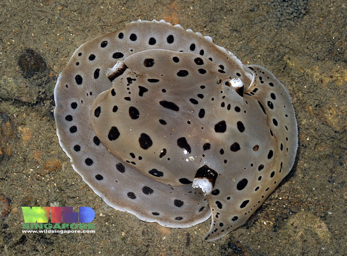
Unlike the many species of nudibranchs that can be found in Singapore, only two species of side-gilled slugs occur locally: Pleurobranchus forskalii and Euselenops luniceps (otherwise known as the moon-headed side-gilled slug). Apart from their drastically different appearances, E. luniceps is also much smaller than P. forskalii, ranging from 3 to 7 cm in length (Tan, 2012a).
 |
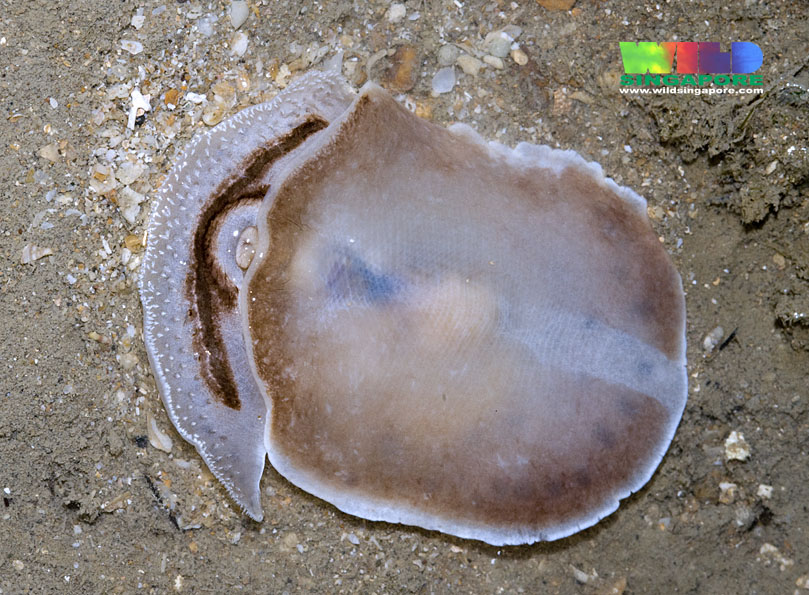 |
| Topside of E. luniceps. Photo credit: Ria Tan. |
Underside of E. luniceps. Photo credit: Ria Tan. |
Biology
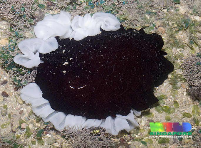 |
| Deposition of egg mass (white ribbons) by P. forskalii. Photo credit: Ria Tan. |
Diet
Members of the genus Pleurobranchus feed exclusively on ascidians (Cimino & Ghiselin, 1999; Cimino et al., 2001), such as Lissoclinum species (Fu et al., 2004), from which they sequester metabolites (see 'Biomedical Importance', under 'Uses') and use in their acid secretions (see 'Defense', this section).Predators
Chitinous jaw plates of P. forskalii have been found in the stomach of a turtle in Tanzania (Rudman, 1999). Currently, no other predators are known for this species.Reproduction
P. forskalii lays an egg mass consisting of white ribbons (pictured).Defense
Without an external shell (the shell is internal in P. forskalii) to give it physical protection, P. forskalii may seem defenseless. However, it is far from it! Brightly-coloured animals such as P. forskalii are usually in some way harmful to predators and their colouration is thus a 'stay away!' warning to potential enemies. This is known as aposematism and it is a type of defense mechanism. For all side-gilled slugs, epidermal cells all over their body secrete a highly acidic mixture containing sulphuric acid (pH 1) that is a very effective deterrent. It is no surprise then that dead specimens are consumed right away (Cimino et al., 2001).Locomotory Behaviours
Crawling
All side-gilled slugs primarily move by crawling. Most of the species crawl using mucociliary locomotion in which the "cilia on the bottom of the foot beat and propel the animal over a surface of secreted mucus" (Newcomb et al., 2012).Swimming
While not yet elucidated for Pleurobranchus forskalii, it is likely to employ the same swimming mechanism as the closely related P. membranaceus. This species swims characteristically via asymmetric undulation, described by Newcomb et al. (2012) as a method of upside down swimming in which the animal uses its mantle "as a passive keel while producing alternating muscular waves along its foot".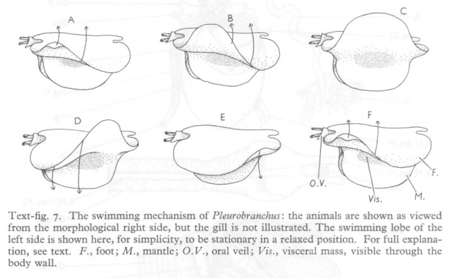 |
| Asymmetric undulation of P. membranaceus during swimming. Extracted from Thompson & Slinn (1959). |
Distribution
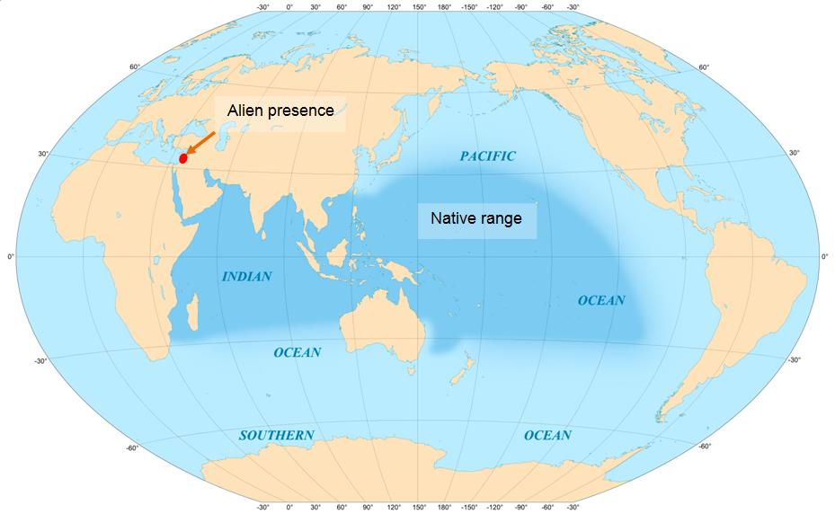
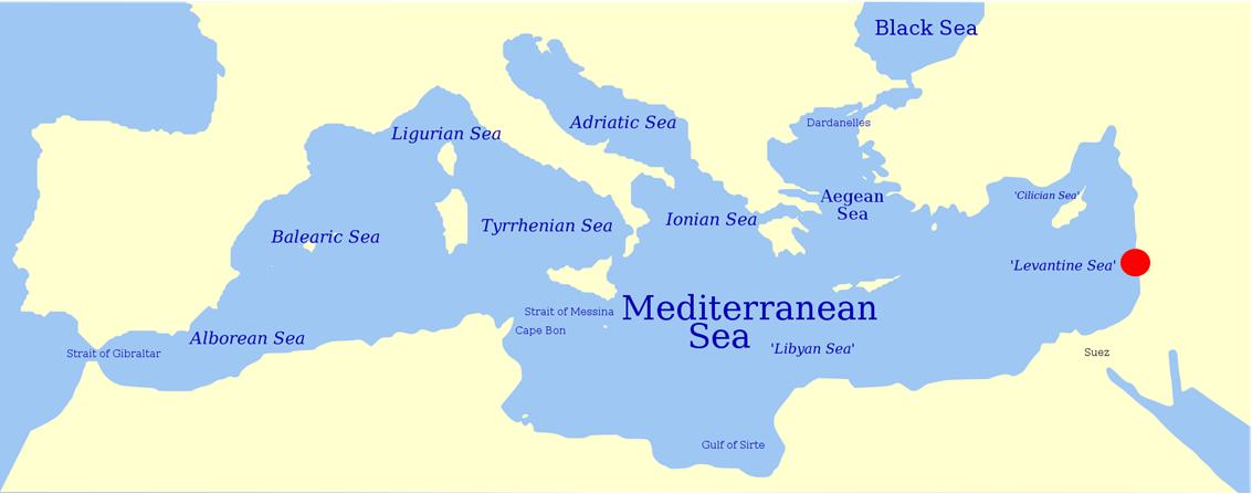
The native range of Pleurobranchus forskalii extends throughout the Indo-Pacific region (Rudman, 1999: top picture). In 1975, the first and only record of P. forskalii as an alien species was made on the Mediterranean coast of Israel (both pictures), at Haifa Bay, with the Suez Canal believed to be its passage of entry (Barash & Danin, 1977). Zenetos et al. (2005) classified the presence of P. forskalii as a 'casual species' – species which have only been recorded once (no more than twice for fishes) in the scientific literature, and are presumed to be non-established in the basin. DAISIE (Delivering Alien Invasive Species Inventories for Europe), however, lists //P. forskalii// as an established alien species.
Uses
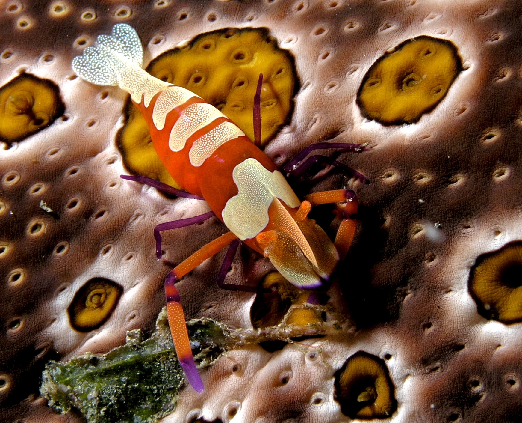 |
| Periclimenes imperator on a sea cucumber host (Bohadschia argus). Photo credit: Nick Hobgood (Wikimedia Commons User: nhobgood). |
Biomedical Importance
P. forskalii contains important metabolites which are sequestered from their diet of ascidians. It is not known if they produce other metabolites on their own (i.e. de novo synthesis). A series of cyclic peptides (Wesson & Hamann, 1996) and chlorinated diterpenes (Fu et al., 2004) have been isolated from P. forskalii. Among the former, keenamide-A exhibits significant activities against the P-388, A-549 and MEL-20 at 2.5 g/mL, and HT-29 at 5.0 g/mL tumor cell lines (Cimino et al., 2001). Two out of the eight diterpenes "were found to be potent cytotoxins in the National Cancer Institute’s screening panel of 60 tumor cell lines and showed some selectivity for melanomas" (Fu et al., 2004).
Threats
In Singapore, Pleurobranchus forskalii is potentially threatened by habitat loss and degradation due to coastal reclamation works and dredging. In addition, suitable habitats for P. forskalii i.e. coral rubble and seagrass are uncommon, so any further impact is likely to reduce its range which may restrict gene flow between (meta)populations.
Conservation Status
Pleurobranchus forskalii has not yet been evaluated for the IUCN (International Union for Conservation of Nature) Red List of Threatened Species.
Morphology
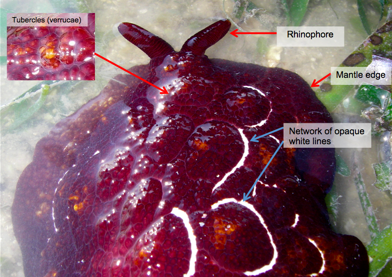 |
| External morphology of P. forskalii. Image courtesy of Toh Chay Hoon (with modifications). |
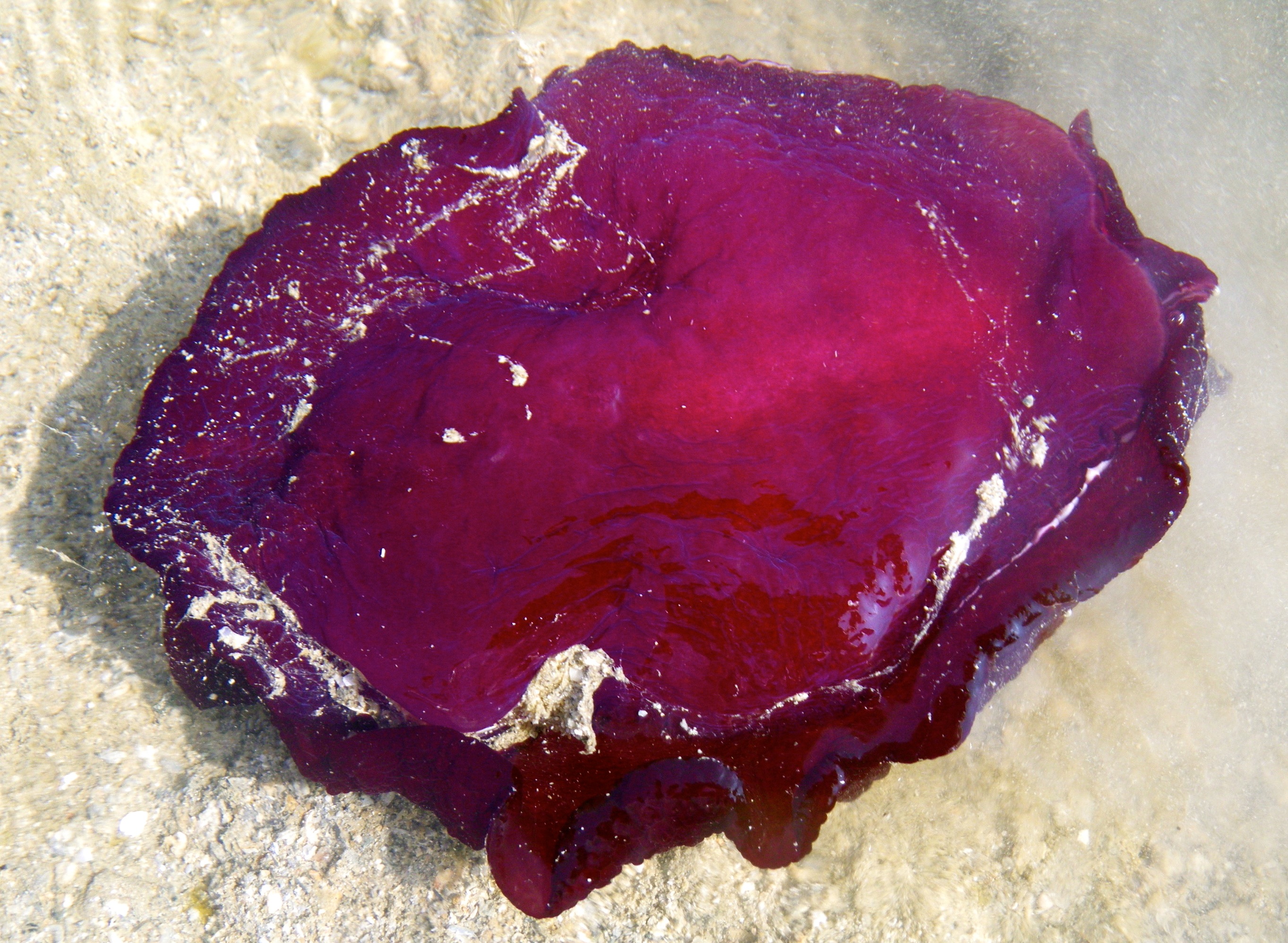 |
| Underside of P. forskalii showing a smooth foot. Own photo (Henrietta Woo). |
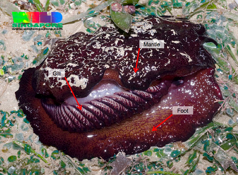 |
| P. forskalii with its mantle flipped over to reveal the gill lying just above the foot. Image courtesy of Ria Tan (with modifications). |
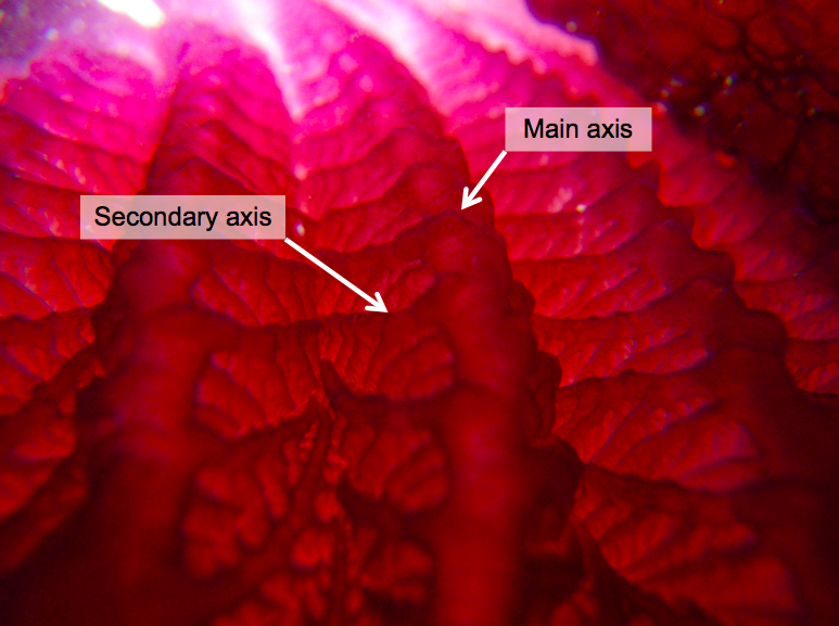 |
| Close-up of main and secondary gill axes. Own photo (Henrietta Woo) with modifications. |
| Proboscis of P. forskalii with its tip retracted. Own photo (Henrietta Woo). |
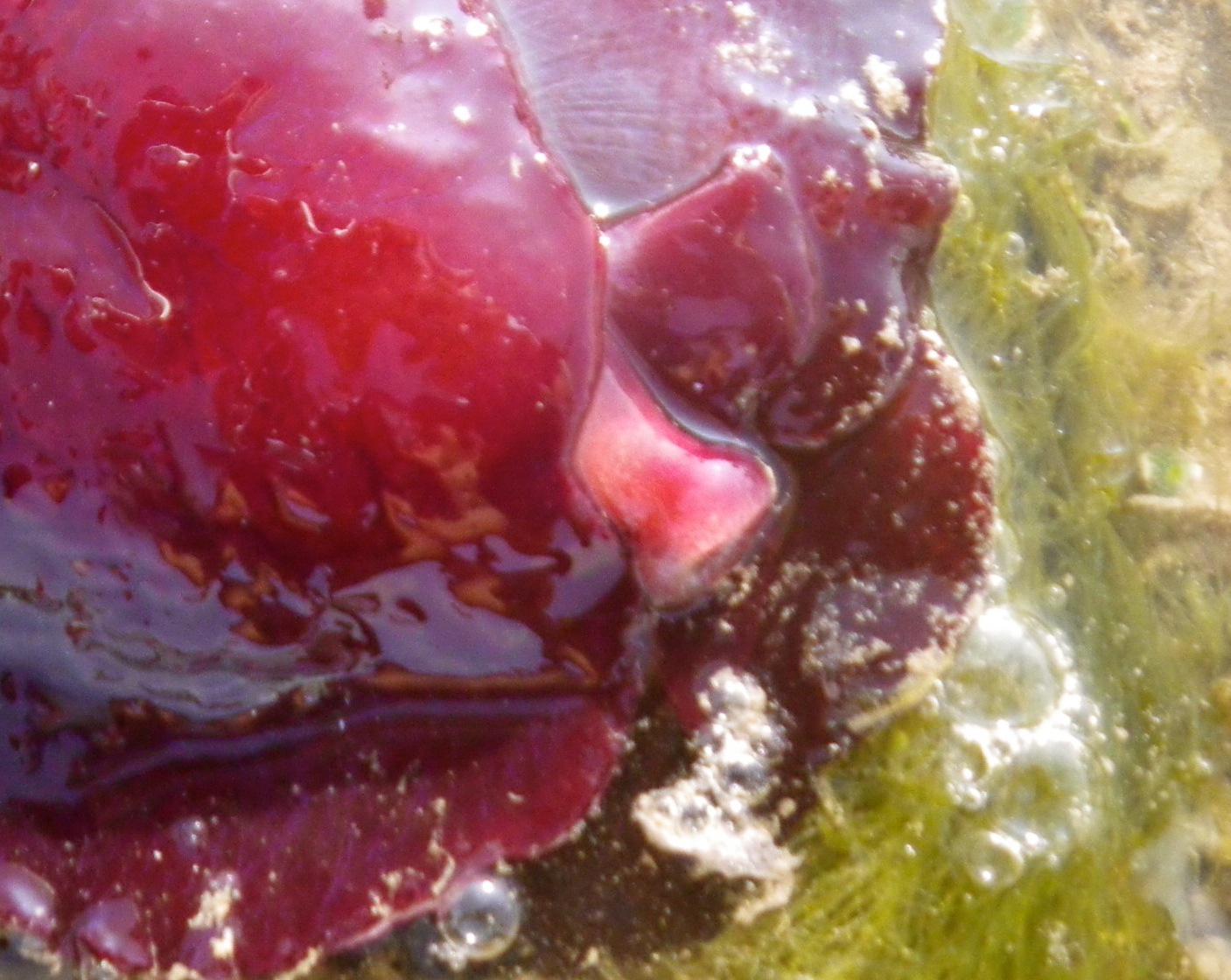 |
| Proboscis of P. forskalii with its tip expanded. Own photo (Henrietta Woo). |
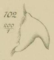 |
| Drawing of the hook-shaped first tooth of the radula of P. forskalii. Extracted from Vayssière (1898). Image courtesy of Biodiversity Heritage Library. http://www.biodiversitylibrary.org. |
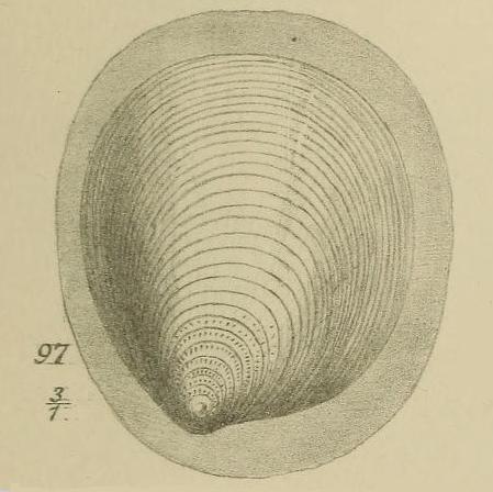 |
| Internal shell of P. forskalii. Extracted from Vayssière (1898). Image courtesy of Biodiversity Heritage Library. http://www.biodiversitylibrary.org. |
The shell of P. forskaliiis located nearer to the head of the animal rather than its rear, and is described as being small and rounded. It appears to be quite membraneous, transparent and thin (Vayssière, 1898).
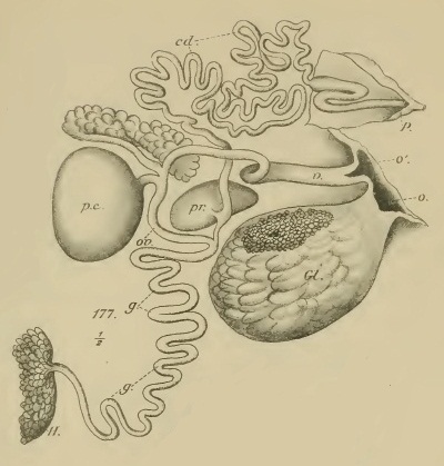 |
| Reproductive system of P. forskalii. Extracted from Vayssière (1898). Image courtesy of Biodiversity Heritage Library. http://www.biodiversitylibrary.org. |
For most, if not all, sea slugs, individuals have both male and female reproductive organs in their body i.e. they are hermaphrodites. Here, the reproductive system of P. forskalii is depicted and the labelled parts are as follows: H, hermaphrodite gland; g, g, genital tract common; pr, prostate; cd, cd, vas deferens; p, penis; ov, ov, oviduct; pc, pocket copulatrix; v, vaginal region of the oviduct with its orifice external; o', Gl, clusters formed by mucus glands and albumin; o, external orifice recent glands.
Original Description
Original descriptions of species are important in taxonomy as it provides an undisputed source of reference should doubt over the original designation of the species arise. The description is based on a type specimen (or sometimes a series of specimens) which serves as a "scientific name-bearing representative for any animal or plant species" (Smithsonian National Museum of Natural History, 2008). Though several different kinds of type specimens exist, the most common is the holotype. The holotype is kept in a public depository such as a museum and can subsequently be examined by taxonomists.
Based on the original description of Pleurobranchus forskalii by Rüppell & Leuckart, [1828], the type specimen is deposited in Museo Francoforte i.e. the Museum of Frankfurt (the Senckenberg Museum) and the type locality i.e. where it was originally found is given as Massawa, a city located on the Red Sea coast of Eritrea. An English version of the original description is given below, translated from German/Latin/French/Italian using Google Translate. Where the translation was far from being understandable, I have edited it at my own discretion or left it in the original language. Any errors are completely my own. Note that the name 'Lepus marinus' mentioned below is a vernacular and has traditionally been applied to sea hares as well as similar gastropods (Rudman, 1993).
| Original description of P. forskalii (page 18). Extracted from Rüppell & Leuckart, [1828]. Image courtesy of Biodiversity Heritage Library. http://www.biodiversitylibrary.org. |
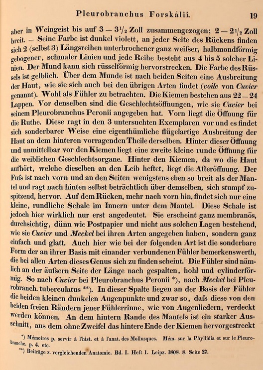 |
| Original description of P. forskalii (page 19). Extracted from Rüppell & Leuckart, [1828]. Image courtesy of Biodiversity Heritage Library. http://www.biodiversitylibrary.org. |
Gen. Pleurobranchus. (Cuv.)
Table 5
(Fig. 2 a, b)
Pleurobranchus Forskalii
(Mus. Francof.)
Diagnosis. Pleurobranchus. The dorsal surface of the body is rough, coloured a dark violet. There are a series of two (or three) discontinuous lines, shaped like a semi-crescent, on the dorsal surface of the animal. An incision on the posterior mantle is noted.
The habitat of P. forskalii is in the Red Sea, near Massawa.
Forskal *) this species has been rather well and clearly illustrated by the name Lepus marinus, but nowhere described. Later zoologists have left the figure usually unnoticed. Only when Lamarck ) we find that the same sex is detected correctly, but in an incorrect way, as though in question, that Thier reckoned to Pleurobranchus peronii. According to Cuvier *): "The figure of Forskal, P. XVIII. is probably Pleur. Peronii."
*) Icons of Nature of the Eastern Routes Depicting P. Forskal etc. Havniae 1776. Tab. XXVIII. A.
) The Natural History of Animals Without Veterbrates. Tom. VI. Part. 1. p. 339.
*) The Animal Kingdom. Tom. II. p. 396. Note 1.
This species that we first describe is undoubtedly the giant of all hitherto known Pleurobranchen and in life 5 to 6 inches long, but in spirit it contracted to 3 to 3.5 inches, and 2 to 2.5 inches wide. Its color is dark purple, and on each side of the back there are 2 (even 3) longitudinal rows of interrupted completely whiter, crescent curved, narrow lines and each line consists of 4 to 5 such lines. The proboscis may stretch out of the mouth into a snout. The color of its trunk is yellowish. About the mouth on both sides are spreading skin as they are also found in other species (called veil of Cuvier). Probably to be considered as a sensor. The gills consist of 22 to 24 lobes. Before the same are the sexual openings, as Cuvier has given in his description of Pleurobranchus peronii. At the front is the opening for the penis. This protrudes outside of the study in the three tested specimens, and there is a peculiar winglike spreading of the skin at the rear protruding part thereof. Behind this opening and just before the gills is a second small round opening for the female reproductive organs. Behind the gills, where the skin stops, which attaches the same way to the body, is the anus. The foot continues to the front and on the sides it is at least as wide as the shell and extends to the rear even considerably over the same to produce blunt tapering. On the back, more towards the front, only a small, plump shell inside is found under the mantle. This shell is really only hinted at before. It appears quite membranous, translucent, thin as paper mail, not based on such documentation as Cuvier and Meckel gave for their species, but simple and smooth. Here too, as in the following way is the singular form of the head at its base with interconnected rhinophores installed in all species of this genus. The rhinophores are in fact on the side longitudinally split hollow and are cylindrical. Thus, according to Cuvier at Pleurobranchus peronii *), according to Meckel at
| Original description of P. forskalii (page 20). Extracted from Rüppell & Leuckart, [1828]. Image courtesy of Biodiversity Heritage Library. http://www.biodiversitylibrary.org. |
Found on coral at Massawa in January.
*) Mémoire p. servir à l'hist. et à l'anat. des Mollusques. Mém. sur la Phyllidia et sur le Pleurobranche. p. 4. etc.
) Contributions to Comparative Anatomy. Vol I Issue 1 Leipz. 1808. 8. Page 27.
| Fig. 2a from the original description of P. forskalii. Extracted from Rüppell & Leuckart, [1828]. Image courtesy of Biodiversity Heritage Library. http://www.biodiversitylibrary.org. |
| Fig. 2b from the original description of P. forskalii. Extracted from Rüppell & Leuckart, [1828]. Image courtesy of Biodiversity Heritage Library. http://www.biodiversitylibrary.org. |
Taxonavigation
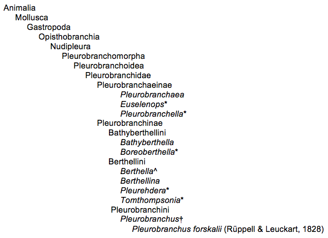
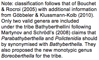
*monotypic genera
†largest pleurobranchomorphan genus
^second largest pleurobranchomorphan genus
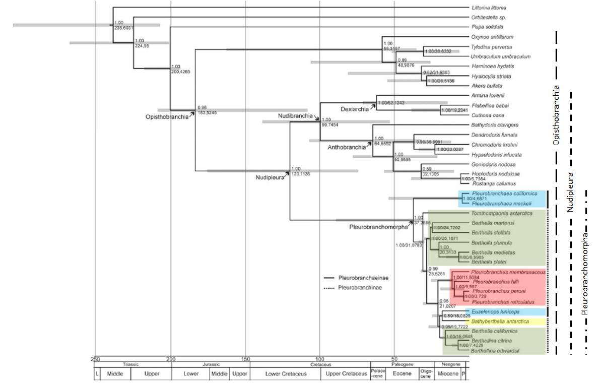 |
| Maximum clade credibility tree displaying results of the relaxed molecular clock analyses. Posterior probabilities and age estimations (in Million years) are provided at the branches. Grey node bars indicate 95% HPD (highest posterior density) credible intervals. Geological time scale is based on the International Stratigraphic chart by the International Commission on Stratigraphy (2008). (L, Lower; P, Pilocene). (Blue - Pleurobranchaeinae; green - Berthellini; red - Pleurobranchini; yellow - Bathyberthellini). Modified from Göbbeler & Klussmann-Kolb (2010). |
The Pleurobranchomorpha comprise the side-gilled slugs which are a sister group to the Nudibranchia or nudibranchs. Together, they make up the Nudipleura (Wägele & Willan, 2000) – a group that multiple studies (both morphological and molecular-based) have revealed support for (cited in Göbbeler & Klussmann-Kolb, 2010). Though side-gilled slugs were traditionally classified under the Notaspidea, this has since been found to be erroneous given that it is not monophyletic (Schmekel, 1985). Phylogenetic analyses using the four gene markers of 18S rDNA, 28S rDNA, 16S rDNA and CO1 indicate that the divergence of the Nudipleura into the Pleurobranchomorpha and Nudibranchia most probably occurred in the Cretaceous(145 to 66 million years ago; mya) (Göbbeler & Klussmann-Kolb, 2010). The analysis by Göbbeler & Klussmann-Kolb (2010) clearly support an Antarctic origin for the Pleurobranchinae which rapidly radiated during the Oligocene (34 to 23 mya) and early Miocene (approximately 23 to 5 mya), where all main lineagesevolved in a period of about 10 to 15 million years (Göbbeler & Klussmann-Kolb, 2010). About 11.5 mya, radiation of the Pleurobranchini occurred and the species e.g. Pleurobranchus forskalii probably evolved in warm and shallow waters (Martynov & Schrödl, 2008).
Other Species Pages
Gofas, S., G. C. Carrada, J. Templado, A. Zenetos, V. Gollino, 2003. Pleurobranchus forskalii. CIESM: The Mediterranean Science Commission. http://www.ciesm.org/atlas/Pleurobranchusforskalii.html. (Accessed November 2012).
Pittmam, C. & P. Fiene, n.d. Pleurobranchus forskalii. Sea Slugs of Hawai`i. http://seaslugsofhawaii.com/species/Pleurobranchus-forskalii-a.html. (Accessed November 2012).
Rudman, B., 1999. Pleurobranchus forskalii (Rüppell & Leuckart, 1828). Sea Slug Forum. Australian Museum, Sydney. http://www.seaslugforum.net/factsheet/pleufors. (Accessed November 2012).
Tan, R., 2012. Forskal's sidegill slugs (Pleurobranchus forskalii) on the Shores of Singapore. Wild Singapore. http://www.wildsingapore.com/wildfacts/mollusca/slugs/notaspidae/pleurobranchus.htm. (Accessed November 2012).
Wikipedia, 2012. Pleurobranchus forskalii. http://en.wikipedia.org/wiki/Pleurobranchus_forskalii. (Accessed November 2012).
References
Barash, A. & Z. Danin, Z., 1977. Additions to the knowledge of Indo-Pacific Mollusca in the Mediterranean. Conchiglie, 13(5–6): 85–116.
Bouchet, P. & J. P. Rocroi, 2005. Classification and nomenclator of gastropod families. Malacologia, 47(1–2): 1–397.
Cervera, J. L., R. Cattaneo-Vietti & M. Edmunds, 1996. A new species of notaspidean of the genus Pleurobranchus Cuvier, 1804 (Gastropoda, Opisthobranchia) from the Cape Verde Archipelago. Bulletin of Marine Science, 59(1): 150–157.
Cimino, G., M. L. Ciavatta, A. Fontana & M. Gavagnin, 2001. Metabolites of Marine Opistobranchs: Chemistry and Biological Activity. In: Tringali, C. (ed.), Bioactive Compounds from Natural Sources: Isolation, Characterisation and Biological Properties. Taylor & Francis, London and New York. Pp. 557–637.
Colin, P.L. & C. Arneson, 1995. Tropical Pacific Invertebrates: a Field Guide to the Marine Invertebrates Occurring on Tropical Pacific Coral Reefs, Seagrass Beds and Mangroves. Coral Reef Press, California, USA. 296 pp.
Fransen, C. H. J. M. & J. Goud, 2000. Chromodoris magnifica (Quoy & Gaimard, 1832), a new nudibranch host for the shrimp Periclimenes imperator Bruce, 1967 (Pontoniinae). Zoologische Mededelingen, 73(18): 273–283.
Fu, X., A. J. Palomar, E. P. Hong, F. J. Schmitz & F. A. Valeriote, 2004. Cytotoxic lissoclimide-type diterpenes from the molluscs Pleurobranchus albiguttatus and Pleurobranchus forskalii. Journal of Natural Products, 67(8): 1415–1418.
Galil, B. S., 2007. Seeing red: alien species along the Mediterranean coast of Israel. Aquatic Invasions, 2(4): 281–312.
Göbbeler, K. & A. Klussmann-Kolb, 2010. Out of Antarctica? – New insights into the phylogeny and biogeography of the Pleurobranchomorpha (Mollusca, Gastropoda). Molelcular Phylogenetics and Evolution, 55(3): 996–1007.
ICZN Opinion 1767, 1993. Pleurobranchus forskalii Rüppell & Leuckart [1828] and P.testudinarius Cantraine, 1835 (Mollusca, Gastropoda): specific names conserved. Bulletin of Zoological Nomenclature, 51(2): 164–165.
Man and Mollusc, n.d. Gastropoda. http://www.manandmollusc.net/advanced_introduction/moll101gastropoda.html. (Accessed November 2012).
Martynov, A. V. & M. Schrödl, 2008. The new Arctic side-gilled sea slug genus Boreoberthella (Gastropoda, Opisthobranchia): Pleurobranchoidean systematics and evolution revisited. Polar Biology, 32(1): 53–70.
Newcomb, J. M., A. Sakurai, J. L. Lillvis, C. A. Gunaratne & P. S. Katz, 2012. Homology and homoplasy of swimming behaviors and neural circuits in the Nudipleura (Mollusca, Gastropoda, Opisthobranchia). Proceedings of the National Academy of Science of the United States of America, 109(Supplement 1): 10669–10676.
Rudman, W. B. 1993. Case 2838. Pleurobranchus forskalii Rüppell & Leuckart [1828] and P. testudinarius Cantraine, 1835 (Mollusca, Gastropoda): proposed conservation of the specific names. Bulletin of Zoological Nomenclature, 50(1): 16–19.
Rudman, B., 1999. Pleurobranchus forskalii (Rüppell & Leuckart, 1828). Sea Slug Forum. Australian Museum, Sydney. http://www.seaslugforum.net/factsheet/pleufors. (Accessed November 2012).
Rüppell, E. & F. S. Leuckart, [1828]. Neue wirbellose Thiere des Rothen Meers. Pp. 1–22, pls. 1–6. In: Atlas zu der Reise im nördlichen Afrika von Eduard Rüppell. Bronner, Frankfurt-am-Main.
Schmekel, L., 1985. Aspects of Evolution Within the Opisthobranchia. In: Wilbur, K. M. (ed.), The Mollusca. Academic Press, London. Pp. 221–267.
Smithsonian National Museum of Natural History, 2008. What are type specimens? http://collections.mnh.si.edu/whataretypes.html. (Accessed November 2012).
Tan, 2012a. Moon-headed Sidegill Slug (Euselenops luniceps) on the Shores of Singapore. Wild Singapore. http://www.wildsingapore.com/wildfacts/mollusca/slugs/notaspidae/euselenops.htm. (Accessed November 2012).
Tan, R., 2012b. Forskal's Sidegill Slugs (Pleurobranchus forskalii) on the Shores of Singapore. Wild Singapore. http://www.wildsingapore.com/wildfacts/mollusca/slugs/notaspidae/pleurobranchus.htm. (Accessed November 2012).
Tan, S. K. & R. K. H. Yeo, 2010. The intertidal molluscs of Pulau Semakau: preliminary results of "Project Semakau". Nature in Singapore, 3: 287–296.
Thompson T. E. & D. J. Slinn, 1959. On the biology of the Opisthobranch //Pleurobranchus membranaceus//. Journal of the Marine Biological Association of the United Kingdom, 38(3): 507–524.
Vayssière, A. 1896. Description des coquilles de quelques especes nouvelles ou peu connues de Pleurobranchides. Journal de Conehyliologie, 44: 113–137.
Wägele, H. & R. C. Willan, 2000. Phylogeny of the Nudibranchia. Zoological Journal of the Linnean Society, 130(1): 83–181.
Wells, E. E. & C. W. Bryce, 1993. Sea slugs of Western Australia. Western Australian Museum, Australia. 184 pp.
WiIlan, R. C. & N. Coleman, 1984. Nudibranchs of Australasia. Australasian Marine Photographic Index, Australia. 56 pp.
Zenetos, A., M. E. Çinar, M. A. Pancucci-Papadopoulou, J. G. Harmelin, G. Furnari, F. Andaloro, N. Bellou, N. Streftaris & H. Zibrowius, 2005. Annotated list of marine alien species in the Mediterranean with records of the worst invasive species. Mediterranean Marine Science, 6(2): 63–118.
Speak of the Side-gilled Slug
If you have any queries or suggestions on how to improve this page (e.g. you know of new information concerning Pleurobranchus forskalii, spot an error or missing information), do let me know here or at woo.henrietta@gmail.com. Please also use this discussion area to log recent sightings and/or observations of P. forskalii in Singapore, which would greatly contribute to an understanding of its ecology of which precious little is currently known.
comments powered by Disqus
| Subject | Author | Replies | Views | Last Message |
|---|---|---|---|---|
| No Comments | ||||
