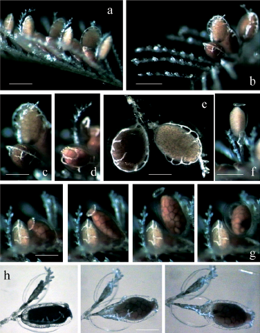
Figure 1. Macrorhynchia philippina: medusoid release. (a) Lateral view of a branch with phylactocarps bearing male and female gonangia. (b) View from above of two phylactocarps with proximal female and distal male gonothecae. (c and d) Details before and during female medusoid release. (c) With a male medusoid still inside the gonotheca, and (d) lacking male medusoid, which wasliberated before the female medusoid; notice the refringent corpuscles. (e) Gonothecae where the ectoderm detaches from the perisarcjust before medusoid release, and with blastostyle plus gubernacula. (f) Profile view of male medusoid issued from the gonotheca, withthe remaining ectodermal sheath bearing blastostyle and gubernacula on one side. (g) Four steps of female medusoid release, from left: engaged, with oocyte mass compressed and extruding through the ring of corpuscles; still engaged; completely free with the bell and theoocyte mass rightly positioned; first contraction inducing the swimming by the apex of the bell. (h) Three steps of the slipping out fromboth the gonotheca and ectodermal sheath of a female medusoid (under an empty male gonotheca). Scale bars a–b, e–f, 1 mm; c–d, g–h, 0.75 mm. All photographs are from video records of living material; a–g, episcopy; h, diascopy and episcopy.

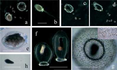
Figure 2. Macrorhynchia philippina: medusoid structure and spawning. (a) Sexual dimorphism of male and female medusoids during spawning. (b) Male expelling spermatozoa during swimming by contraction of the bell. (c and d) Female spawning: a few oocytes expelled during a single contraction of the bell. (e) Female with oocytes detaching from the spadix and mesoglea thickened around the orifice of the bell. (f) Dead empty medusoids with eccentric spadix, white in male and black in female where one oocyte remains stuck. (g) Empty live female medusoid from below, showing bell orifice with velum encircled by a ring of corpuscules, and pigmented cells on the exumbrella and on the velum; insert shows concentrically-arrayed refringent corpuscles after medusoids were desiccated and exposed to hydrogen peroxide. (h) Planula. Scale bars a–f, 1 mm; g–h, 0.5 mm. All photographs are from video records; a–d, f, episcopy; e, diascopy plus episcopy; g, h, diascopy.
From Bourmaud, C., & Gravier-bonnet, N. (2004). Medusoid release and spawning of Macrorynchia philippina Kirchenpauer, 1872 (Cnidaria, Hydrozoa, Aglaopheniidae). Hydrobiologia, 530-531, 365–372. [2]