White stingerMacrorhynchia philippina (Kirchenpauer 1872)
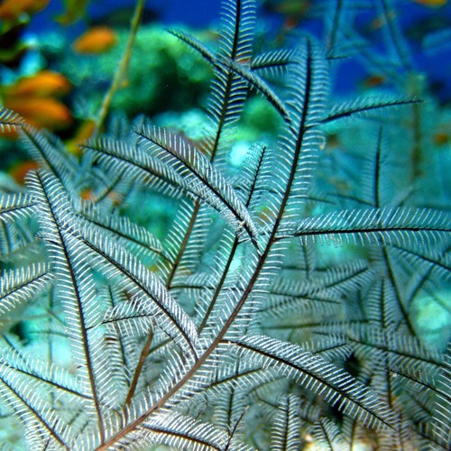
Are you a big fan of diving? Then if you are lucky, you may already have seen this intriguing bush-like animal, growing near coral reefs, in clear and streamy water...
Wondering what it is?
============================================= |
============================================== |
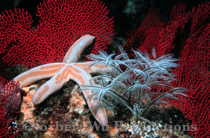 Macrorhynchia philippina in coral reefs. Permission for reproduction pending from Norbert Wu Productions. |
Taxonomic clues:Scientific name: Macrorhynchia philippina (Kirchenpauer 1872) [9] Common names: Stinging bush hydroid, white stinger Synonyms (& genus transfer): Lytocarpus philippina (Pictet 1893), Lytocarpus crosslandi (Ritchie 1907), Aglaophenia urens, Aglaophenia perforata, Aglaophenia philippina (Kirchenpauer, 1872). Classification: Animalia --> Cnidaria ------> Hydrozoa ---------> Leptothecata ------------> Aglaopheniidae ---------------> Macrorhynchia (Here you can see where the 30 species in the genus Macrorhynchia are placed in the tree of life). 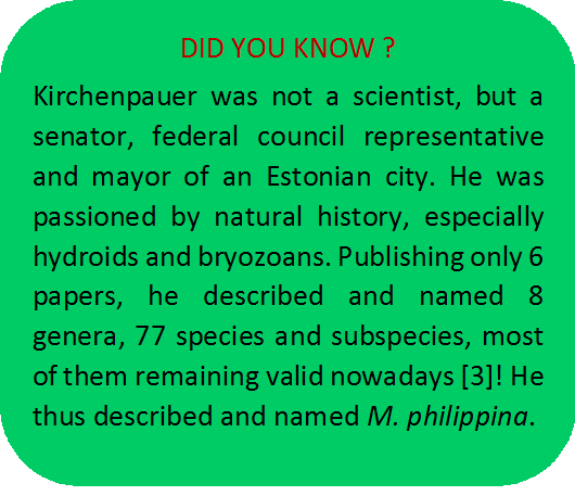 |
Phylogenetics
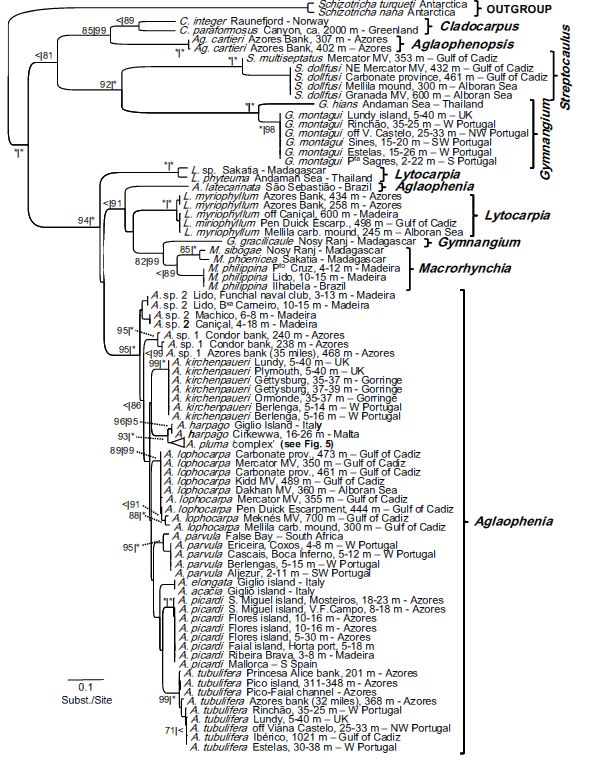
Despite the fact that the family M. philippina belongs to (Aglaopheniidae) is world-wide spread, its phylogeny remains controversial. No proper molecular analyses had been directed until two years ago. Moura et al. ran phylogenetic analyses based on mitochondrial 16S genes sequences. Many of the species studied in that article had never been sequenced so far, hence the authors performed DNA sequencing for the first time for those specimens. They used both maximum likelihood and bayesian methods to retrace the phylogenetic history of the hydrozoan family Aglaopheniidae.
This study focused on mediterranean species. However, Macrorhynchia philippina being progressively invading Mediterranea, Moura et al. also analysed some specimen of our species that they found by hazard in Madeira.
According to this study, the family Aglaopheniidea seems to be monophyletic with high support on the nodes. The clade formed by the genus Macrorhynchia in particular is also well-supported.
Where can you find it?
 M. philippina shows a circumglobal distribution in warm or temperate neritic waters, even if it doesn't occur at high density rates. Indeed, you can find it in the Indian Ocean, Red Sea, East and West Pacific Ocean, East and West Atlantic Ocean, and even in Mediterranea. However, it is interesting to note that it is nowadays considered as an alien species as it has began to spread at unusual latitudes thanks to the global warming [14]. Thus, its migration to the Mediterranean Sea through the Suez Canal shows this invading tendency (species recorded in Turkey [5] and Lebanon [1] for exemple). Restricted to marine environments, this animal cannot tolerate stagnant water in deep bays. Despite the fact that it lives in shallow waters, it does not seem to have lighting preferences since you can find it in places with abundant sunlight as well as at the entrances to caves, where light is less available. World distribution map: http://iobis.org/mapper/?taxon=Macrorhynchia%20philippina |
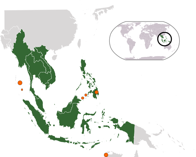 Distribution map of M. philippina in South-East Asia. The orange dots indicate areas where the species has been frequently seen. They are sized according to density of M. philippina populations at the indicated places. Some individuals have been observed in Singapore seas. © ASDFGH - WikiMedia commons. |
How can you recognize it?
General Features:
Macrorhynchia philippina are colonial organisms, that are easily identifiable as iridescent ramified "bushes". Their sizes range from 5 up to 20 centimeters in length and height.
Stalk:The stolons fix the colonies onto the substrate. The main stalk, called hydrocaulus, is brownish-coloured. It bears irregular right-angled ramifications, ramified themselves, giving the colony its bush-shape. The tip ramifications, or hydrocladium, are organized into two alternating ranges all along the stalk. [10] 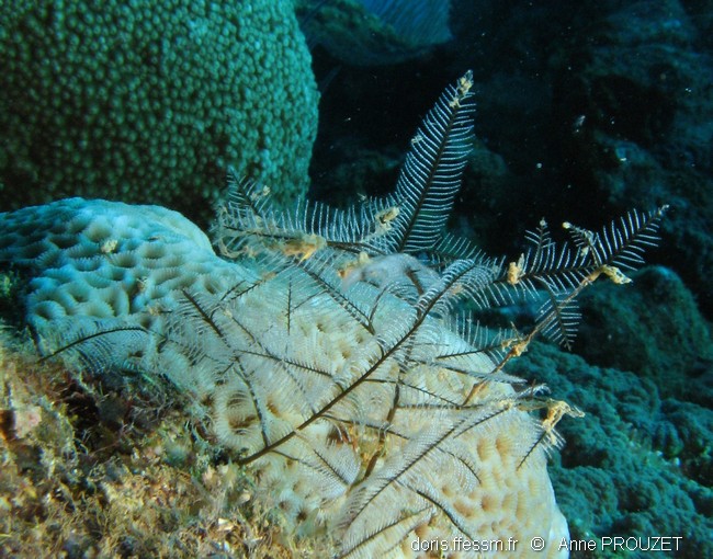 |
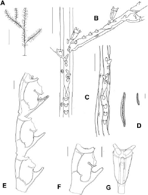 Schemes of M. philippina anatomy: A, colony. B, detail of the hydrocaulus and a hydrocladium. C, basal portion of the hydrocaulus. D, microbasic mastigophores of two sizes. E, hydrocladium in lateral view. F, hydrotheca in lateral view. G, hydrotheca in frontal view. Scale bars: A, 1 cm. B, C, 500 mm. D, 10 mm. E–G, 100 mm. [6] |
 Feather:The hydrocladium are usually of the same length (around 5 centimeters), except at the tips, where they become shorter. M. philippina seems to bear feathers thanks to these features. The feathers are lightly bent and hanging at the tips. |
"Feathers" of M. philippina. © Anne Prouzet 2009 (Published in the DORIS Web site). |
Polype:The hydranths located on hydrocladia are the reason for the white-bluish iridescence of the bush, as they are bright and reflect light. They are surrounded by unstalked hydrothecae (extended from the perisarc), which are aligned on one single row in the direction of the stream. There are three associated nematothecae for each hydrotheca (two lateral and one lower-median). [15] 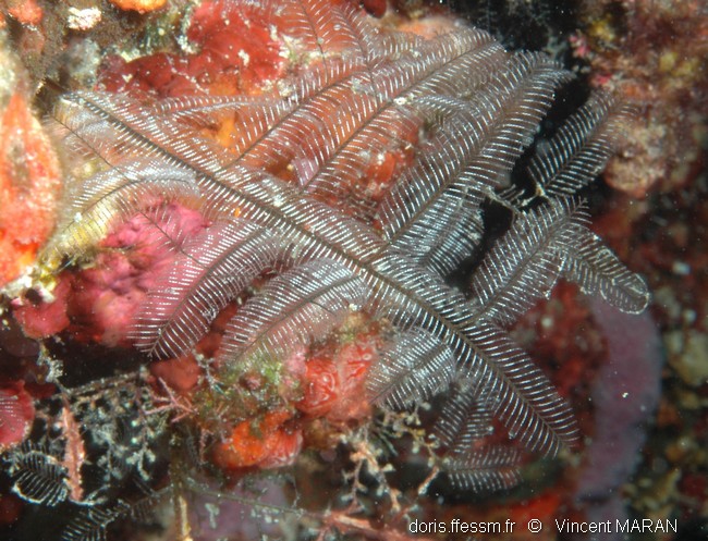 Here, the iridescence of the bush is well-visible. © Vincent Maran 2006 (Published on the DORIS Web site). |
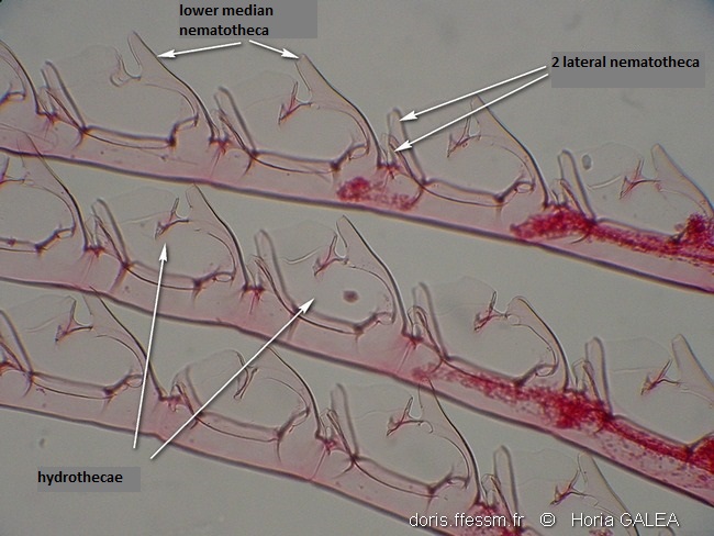 Three portions of cladia with successive hydrothecae. © Horia Galea 2005 (Published on the DORIS Web site). |
How do they live?
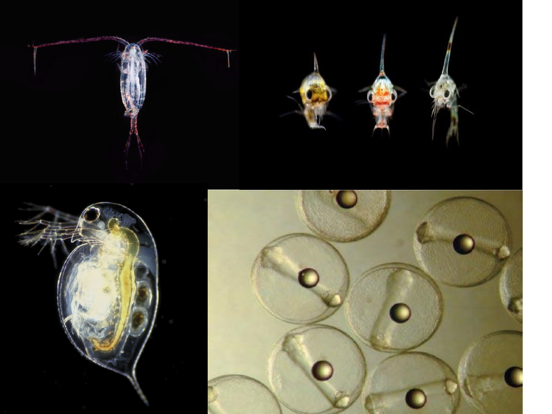 A sample of what M. philippina can feed on. Photos of, from left to right and top to bottom: copepod, crabs larvas, daphnia (© Richard Kirby. Permission for reproduction pending), finfish eggs (© Erik Baard. Permission for reproduction pending). |
Dietary habit:Stinging bush hydroids are active filter-feeders. They capture plankton dwelling in the water currents, with the tentacles of the polyps specialized for feeding (the hydranth).  |
| Reproduction: The reproduction mechanism of M. philippina is not well understand. Although meduseae stage disappeared from the characters of hydroids in the suborder Plumularida , M. philippina appears to be able to undergo both sexual and asexual reproduction [13]. However, sexual reproduction has only been described recently, in a few cases of that species. Typical life cycle: Being hermaphrodites, the white stingers have specialized twigs called phylactocarps. They bear gonozooids at their base, which are wrapped by nematophores for protection [8]. Each phylactocarp contains one female and one male gonotheca, differentiable by colour: red ochre for the female, yellow ochre for the male. The reproductive polyps, also called gonangium, simultaneously release their male and female medusa buds when sexual products are mature [2]. The medusoids then actively swim for about two hours in the water column, disseminating the gametes via contraction of the gonophore bell. The fertilized eggs will grow into a planula larva after 24 hours, which will subsequently either swim or crawl to find a favorable substrate to settle. In hydroids, reproductive events can be influenced by environmental conditions such as temperature, depth, or latitude. 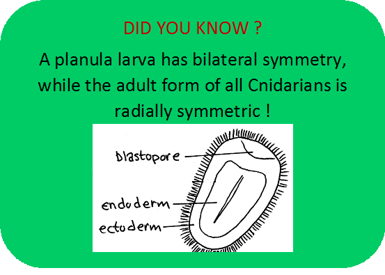 |
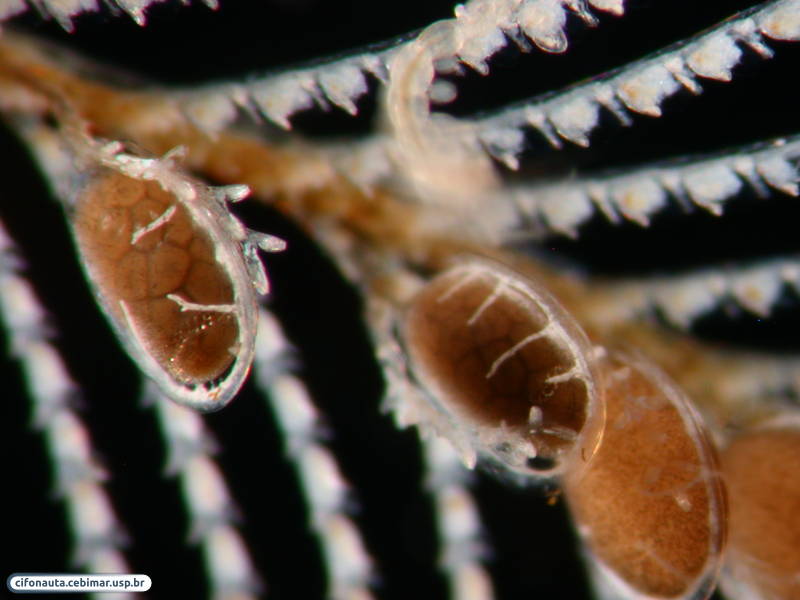 From top to bottom: photograph of a phylactocarp bearing gonangiums. The yellow arrow indicates a gonangium (© Denis Ader 2010. Published on the DORIS Web site). Close view of gonangiums (© Alvaro E. Migotto) Click here to see details of medusoid release, medusoid structure & spawning. |
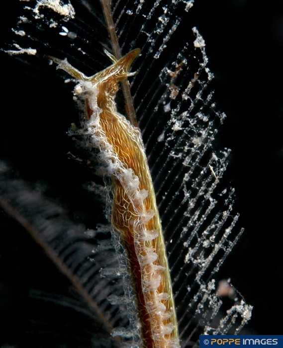 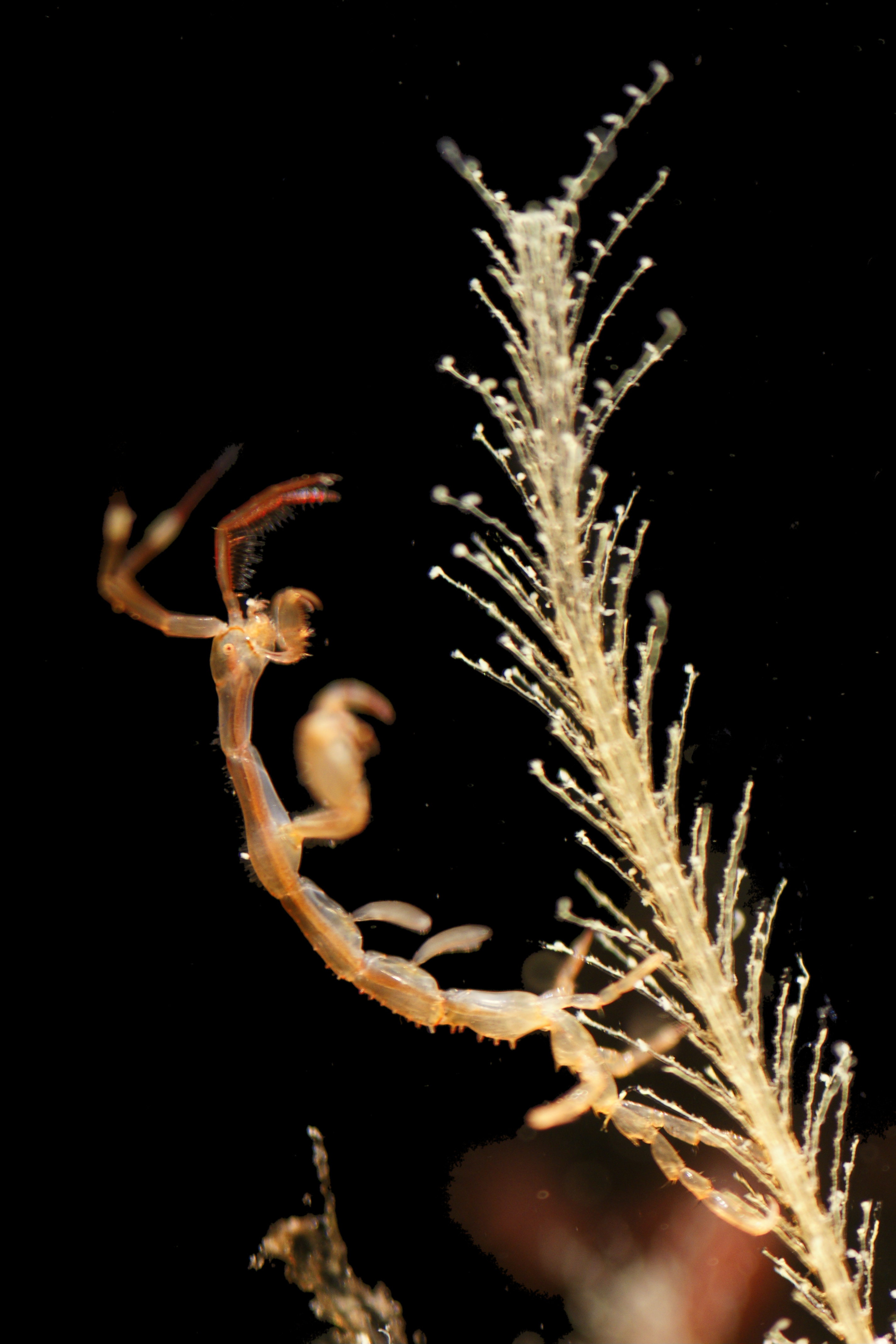 Photo of (from left to right): L. vermiformis feeding on M. philippinq (© Philippe & Guido Poppe - www.poppe-images.com); ghost shrimp Caprella mutica hiding in the bush formed by the ramifications of a hydroid (© Hans Hillewaert - Wikimedia commons). |
Biological interactions:Predation The urticating cells of the Macrorhynchia philippina that are the source of its common name "stinging hydroid" protects it from most predators. Despite its extremely efficient defenses, M. philippina still have a predator, a mollusc from the Nudibranchia order: Lomanotus vermiformis. This gastropod isn't sensitive to urticating propensities of our hydroid. [16] Parastism Stinging bush hydroids are host to several ectoparasites, all from the same genus: Macrochiron. These parasites are Crustaceans from the Copepoda subclass. They live on and consume the mucus released by M. philippina. [17] Commensalism The ramified bushes provide refuge for ghost shrimps to hide. |
Why are they important?
In Biology:
"Rare events contain more information" (Information Theory, Shannon, 1948).
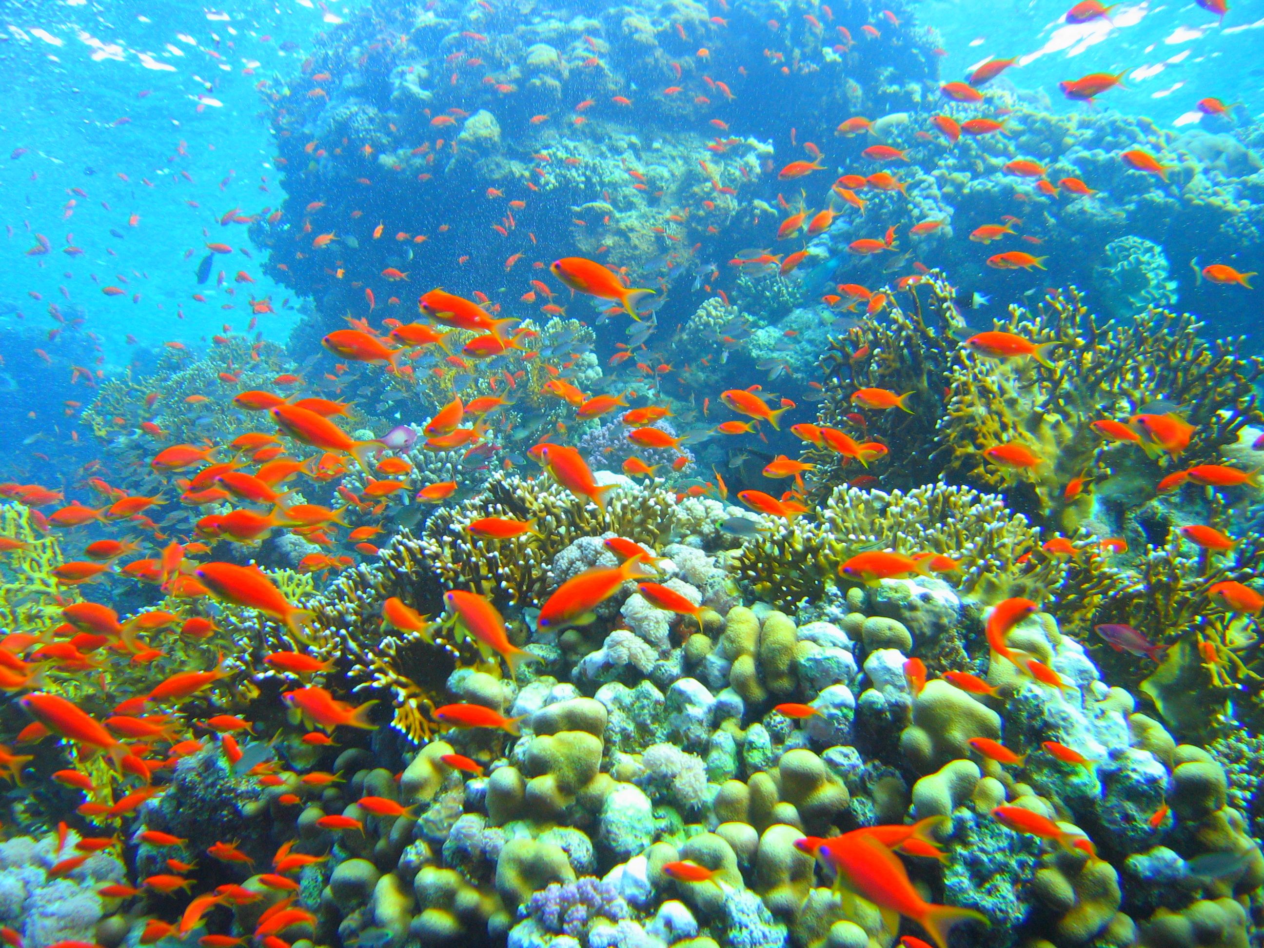 This probabilistic statement can be applied in biodiversity and conservation biology. As an uncommon species living in the most diverse habitats that are coral barriers [4,7], the presence of Macrorhynchia philippina brings a great information about coral reefs biodiversity and health. It could be used as environment indicators for conservation purposes. Moreover, it is the site of many biological interactions as seen above (predation, parasitism, commensalism). However, M. philippina is not well known, not even much studied, despite of possibly interesting co-evolution processes for example.
 [11]Coral reefs © Mikhail Rogov - Wikimedia Commons.In Medicine & Pharmacology:
The urticating property of Macrorhynchia philippina may become the basis of venom or poison study, and thereby could lead to new therapeutic treatements against poisoning. Protein and DNA study may be important in this perspective. However, there is almost no knowledge about M. philippina at the molecular level.
To see the only partial sequence available for the species (16S ribosomal DNA gene), click here.
For Amateurs:
This is simply a handsome and mysterious animal, that looks like a plant. In addition, it even provides shelter for other organisms! It belongs to one of most wide-spread Cnidarian class, Hydrozoans, that has not been represented on the wikispecies yet. However, be careful when diving not to touch it, or you will experience the stinging properties of M. philippina!
Glossary:
(To go back to the related paragraph, simply click on the word you want.)
Animalia. Kingdom. Heterotrophy, locomotion.
Cnidaria. Phylum. Aquatic, radial symmetry, cnidocytes, polype and/ or medusa stages, larva Planula.
Hydrozoa. Class. Velum, ectodermal gonads, secondary loss of septa and endodermic cnidocytes.
Leptothecata. Order. Theca (chitinous envelop), flattened medusa with statocysts on the ridges of the umbrella, gonads on the radiating canals.
Aglaopheniidae. Family. For more informations, see here: Aglaopheniidae family details
Neritic. Shallow part of the ocean spreading from the low-tide to the boundaries of the continental shelf (200 meters in depth).
Stolon. Extension of the hydrozoan body wall, developping buds that give rise to new polyps. The polyps usually remain connected via the stolon.
Hydranth. Feeding zooid (polype) that bears the mouth and tentaclesand contains the stomach region.
Iridescence. Optical property of certain surfaces that change in colour as the angle of view or the angle of illumination changes. It is most of the time due to structural coloration, that is to say regular microstructures that interfere with light.
Hydrotheca. Extension of the perisarc that surrounds and protects the hydranth when it is contracted.
Perisarc. The outer tegument of a hydroid. Often chitinous.
Nematotheca. small stemmed structures from which nematophores develop.
Phylactocarp. Branch of a plumularian hydroid modified to protect the gonothecae.
Gonozooid. Polyp of the colony specialized in reproduction function. Synonym: gonangium.
Nematophore. Defensive organ of the nematozooids (polyps involved in defense function in the colony).
Medusoid. Shortlived structure devoted to gamete dispersal. Meduse-like system that lacks tentacles, bulbs, circular & radial canals, mouth & sense organs.
Gonophore. Reproductive organ of the gonozooids.
Nudibranchia. Order. Mollusc gastropod."Nacked gills".
Commensalism. Biological interaction from which one organism benefits, without beeing either beneficial, nor harming for the second organism.
Links to others pages about M. philippina:
World Register of Marine Species
DORIS, 27/3/2014 : Macrorhynchia philippina Kirchenpauer, 1872
Sealife Base
References:
[1] Bitar, G. (2010). Impact des changements climatiques et des espèces exotiques sur la biodiversité et les habitants marins au Liban. Rap. Comm. Int. Mer Médit., 39.
[2] Bourmaud, C., & Gravier-bonnet, N. (2004). Medusoid release and spawning of Macrorhynchia philippina Kirchenpauer, 1872 (Cnidaria, Hydrozoa, Aglaopheniidae). Hydrobiologia, 530-531, 365–372.
[3] Calder, D. R., & Brinckmann-voss, A. (2011). Gustav Heinrich Kirchenpauer (1808–1887) of the City of Hamburg, and his research on hydroids and bryozoans. Zootaxa, 59, 49–59.
[4] Calder, D.R. (1997). Shallow-water hydroids of Bermuda : superfamily Plumularioidea, Royal Ontario Museum Life Sciences Contributions, 161, 1–84.
[5] Çinar, M. E., Bilecenoglu, M., Öztürk, B., & Can, A. (2006). New records of alien species on the Levantine coast of Turkey. Aquatic Invasions, 1(2), 84–90.
[6] Di Camillo, C. G., Puce, S., & Bavestrello, G. (2009). Macrorhynchia species (Cnidaria: Hydrozoa) from the Bunaken Marine Park (North Sulawesi, Indonesia) with a description of two new species. Italian Journal of Zoology, 76(2), 208–228.
[7] Gibbons, M. J., & Ryland, J. S. (1989). Intertidal and shallow water hydroids from Fiji. I. Athecata to Sertulariidae. Memoirs of the Queensland Museum 27:377-432.
[8] Gravier-Bonnet, N. (2004). Hydroid nematophores: morphological, structural, and behavioural variety from old knowledge and new data. Hydrobiologia, 530-531(1-3), 199–208.
[9] Kirchenpauer, G. H. (1872). Ueber die Hydroidenfamilie Plumularidae, einzelne Gruppen derselben und ihre Fruchtbehälter. Abhandlungen Aus Dem Gebiete Der Naturwissenschaften Herausgegeben von Dem Naturwissenschaftlichen Verein in Hamburg, 5(3), 1–52.
[10] Millard, N.A.H. (1975). Monograph on the Hydroida of southern Africa, Annals of the South African Museum, 68, 1-513. (under the name Lytocarpus philippinus)
[11] Morri, C., Puce, S., Bianchi, C.N., Bitar, C., Zibrowius, H., Bavestrello, G. (2009). Hydroids (Cnidaria: Hydrozoa) from the Levant Sea (mainly Lebanon), with emphasis on alien species. Journal of the Marine Biological Association of the United Kingdom, 89, 49-62.
[12] Moura, C. J., Cunha, M. R., Porteiro, F. M., & Rogers, A. D. (2012). A molecular phylogenetic appraisal of the systematics of the Aglaopheniidae (Cnidaria: Hydrozoa, Leptothecata) from the north-east Atlantic and west Mediterranean. Zoological Journal of the Linnean Society, 164(4), 717–727.
[13] Nutting, C.C. (1900). American hydroids. Part 1 : The Plumularidae, Special bulletin - United States National Museum, 4 (1), 1-285.
[14] Öztürk, B. (2010). Status of alien species in the Black and Mediterranean Seas. Studies and Reviews. General Fisheries Commission for the Mediterranean, 87. Rome, FAO.
[15] Proceedings of the Biological Society of Washington, (1943), 96.
[16] Rudman, W.B. (1999). Lomanotus vermiformis Eliot, 1908, Sea Slug Forum, Australian Museum, Sydney.
[17] Schuchert, P. (2014). Macrorhynchia philippina Kirchenpauer, 1872, Schuchert, P. (2014) World Hydrozoa database. Accessed through: World Register of Marine Species
Images References
Macrorhynchia philippina in coral reefs. http://www.norbertwu.com/lightbox/detail/724.html
Close-up photo of M. philippina. http://fotki.yandex.ru/users/diving-lv2006
Distribution map of M. philippina in South-East Asia; Ghost shrimp Caprella mutica hiding in the bush formed by the ramifications of a hydroid; Coral reefs. Wikimedia Commons
M. philippina fixed on a coral; "Feathers" of M. philippina; Three portions of cladia with successive hydrothecae; Iridescence of the bush; Photograph of a phylactocarp bearing gonangiums. PROUZET Anne, GALEA Horia, in : DORIS, 27/3/2014 : Macrorhynchia philippina Kirchenpauer, 1872, http://doris.ffessm.fr/fiche2.asp?fiche_numero=1761
A sample of what M. philippina can feed on. http://www.planktonpundit.org/ ; https://naturecalendar.wordpress.com/
Close-view of gonangium. http://www.medslugs.de/E/Photographers/Alvaro_E_Migotto.htm
Scheme of typical planula larva. http://lanwebs.lander.edu/faculty/rsfox/invertebrates/aurelia.html
L. vermiformis feeding on a M. philippina. http://www.poppe-images.com/?t=17&photoid=929302